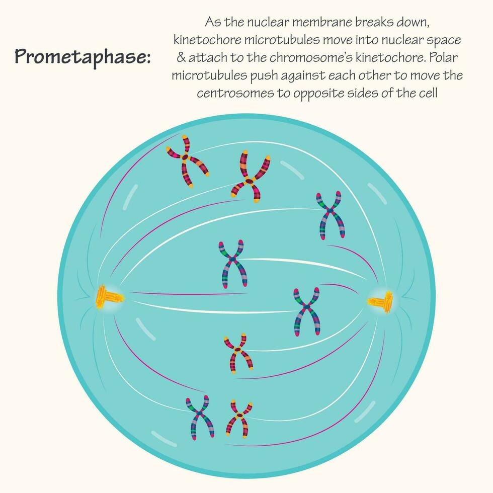
Prometaphase Biology Diagrams
Prometaphase Biology Diagrams The mitosis stages diagram provides a visual representation of the sequential phases of mitosis: prophase, prometaphase, metaphase, anaphase, and telophase. Each stage is characterized by distinct changes in chromatin organization, nuclear envelope breakdown, chromosome alignment, and spindle fiber formation.

Figure 6.4 Animal cell mitosis is divided into five stages—prophase, prometaphase, metaphase, anaphase, and telophase—visualized here by light microscopy with fluorescence. Mitosis is usually accompanied by cytokinesis, shown here by a transmission electron microscope. (credit "diagrams": modification of work by Mariana Ruiz Villareal; credit "mitosis micrographs": modification of work by Prometaphase is the second phase of mitosis, when the nuclear envelope breaks down and kinetochore microtubules attach to the sister chromatids. Learn more about the process and see a diagram of prometaphase at Scitable, a science learning platform. Provide mitosis diagrams for the stages of mitosis; Give you five resources for learning more about the phases of mitosis; Now, let's dive in! After prometaphase ends, metaphase—the second official phase of mitosis—begins. (Kelvinsong/Wikimedia Commons) Phase 2: Metaphase.

Prometaphase Biology Diagrams
Learn about the stages and mechanisms of mitosis, the process of nuclear division in eukaryotic cells. See diagrams of chromosome condensation, spindle formation, and kinetochore attachment.

1.2 Prometaphase 1.3 Metaphase; 1.4 Fig 2 - Summary diagram showing the stages of mitosis. Clinical Relevance - Errors of Mitosis. Errors in mitosis typically occur during metaphase. Usually, this is due to a misalignment of chromosomes along the metaphase plate or a failure of the mitotic spindles to attach to one of the kinetochores

Khan Academy Biology Diagrams
Prometaphase is a phase of mitosis in eukaryotic cells where the nuclear membrane breaks and the chromosomes form kinetochores. The kinetochore microtubules attach to the kinetochores and the chromosomes move to the centre of the cell. This diagram provides a visual representation of the different phases and helps in comprehending the complex process. It also enables scientists and students to identify and recognize the key features associated with each stage. The stages of mitosis include prophase, prometaphase, metaphase, anaphase, and telophase. Learn about the phases of mitosis and their significance in cell division on Khan Academy.
